Eximer Ophthalmology Clinic is equipped with a set of modern high-precision diagnostic facilities, allowing us to determine the functional and anatomical features of the visual apparatus in one visit: visual acuity, intraocular pressure, state of the eye inner structures, retina, and optic nerve, corneal shape, and refracting power, visual fields and many others. The latest certified equipment allows measuring the necessary parameters with no pain or contact. Vision diagnostics is performed according to all world standards.
The diagnostic complex of Eximer Ophthalmology Clinic
Tonoref II Autorefractor (Nidek, Japan)
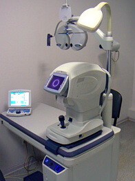
This multifunctional complex includes such devices as an autorefractometer, autokeratometer and non-contact tonometer, which allows performing several types of examinations at once: measuring eye refraction and detecting its disorders (myopia, hyperopia, astigmatism, and their degree); measuring the corneal curvature radius, pupil diameter, and distance between the pupils of the eye. Intraocular pressure is measured without contact using a targeted air jet. Binocular vision and color perception, as well as contrast sensitivity, can be checked.
Echoscan (Nidek, Japan)
This device is required to visualize the shape and properties of the eye’s internal structure. The dimensions of the anterior ocular chamber, lens thickness, eyeball, and vitreous body dimensions can be determined. These data are also used for a variety of microsurgical procedures, including cataract and excimer laser correction.
IOL-master optical coherence biometer (Zeiss, Germany)

It measures the eye length, corneal curvature, and anterior chamber depth at the same time. One of its main functions consists in estimating parameters of the implanted intraocular lens before performing cataract and refractive lens substitution surgeries. These estimates are critical for implanting high-tech lenses with complex optics to maximize visual performance.
Automated perimeter (visual field analyzer) (Zeiss, Germany)

It is used to obtain information about the visual field – the space visible by the human eye at a fixed state. The visual field is affected by diseases of the neuroreceptor apparatus, including the retina and optic nerve (e.g., glaucoma, macular dystrophy). By checking the visual field, you can timely detect the disease and begin the necessary treatment, preventing irreversible vision loss.
EM-3000 Endothelial Microscope (Tomey, Japan)

This equipment determines the quantitative and qualitative composition of the endothelial cell layer of the cornea that ensures its transparency. The analysis and monitoring of the corneal endothelium condition is required before deciding to perform microsurgical procedures in patients with corneal pathology or people wearing contact lenses.
RTVue-100 optical coherence tomography scanner (Optovue, USA)

It produces 2D and 3D images of the retina and the optic nerve disc structures, controls the state of the macula – the central area of the retina responsible for the direct vision, studies the structures of the anterior chamber of the eye. It allows examining the eye ground in patients with initial opacities of the ocular media. Optical coherence tomography provides a precise diagnostic of glaucoma, including assessment of the disease progression and effectiveness of glaucoma treatment. Research with an optical coherence tomography is a priceless aid for specialists in the diagnosis and dynamic monitoring of age-related maculodystrophy, as well as in the case of suspected retinal pathologies. The equipment enables storing the information on each patient in the database.
Orbscan tomography (Bausch&Lomb, USA)
The equipment allows to analyze and evaluate the anterior and posterior surfaces of the eye cornea, the thickness of the cornea over the entire area, the state of the anterior chamber, its central height, angle, and volume, to define quantitatively the optical density of the lens. These examinations are also needed for refractive surgeries, cataract surgery, and glaucoma treatment.
Wave Scan Aberrometer (AMO, USA)

This unique diagnostic device allows determining all visual distortions (aberrations) of lower (myopia, hyperopia, and astigmatism) and higher orders (coma, distortion, spherical aberrations). The data obtained from the aberrometer is used in the course of LASIK Custom Vue personalized laser vision correction.
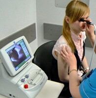
PlusOptix A09 pediatric autorefractometer (Plusoptix, Germany)

This is a unique device to measure eye refraction in children of any age (including infants), children with nystagmus, and challenging patients. It measures the pupil diameter of both eyes and the distance between them, without contact, from a distance of one meter, obtaining a pattern of fixation behavior. All measurements with the PlusOptix A09 autorefractometer are performed in a playful way: the camera emits a special sound, thus attracting the child’s attention and the screen shows a smiling face. It takes just a few seconds to determine the refraction without contact, so the child does not get tired or feel any discomfort.
ARGOS® Biometer with Image Guidance (Alcon)

The ARGOS Biometer facilitates faster preoperative measurements than other market-leading biometers. This reduces the risk of measurement variability and increases the efficiency of cataract planning.The biometer measures data such as:
• Anterior chamber depth (ACD)
• Keratometry (К)
• Corneal diameter (CD)
• Central corneal thickness (CCT)
• Axial length (AL)
• Aqueous humour depth (AD)
• Pupil dimension (PD)
• Lens thickness (LT)
Eximer Ophthalmology Clinic diagnostic facilities include multiple ophthalmoscopes, electronic tonographs, computerized phoropters, and other equipment.
Depending on the findings, an ophthalmologist gives recommendations, makes a treatment program if necessary, and selects the best way to solve your vision problems.