

The SCHWIND SIRIUS+ optical corneal topographer is a system for corneal topographic and aberrometric analysis of the anterior eye segment — a modern diagnostic device that, in a single measurement, provides comprehensive data on the state of the cornea: its thickness, shape, refractive power, as well as all optical distortions (aberrations) of the eye — both lower-order (myopia, hyperopia, astigmatism) and higher-order (coma, distortion, spherical aberration). The accuracy of this data determines the safety and predictability of the outcome of laser vision correction.
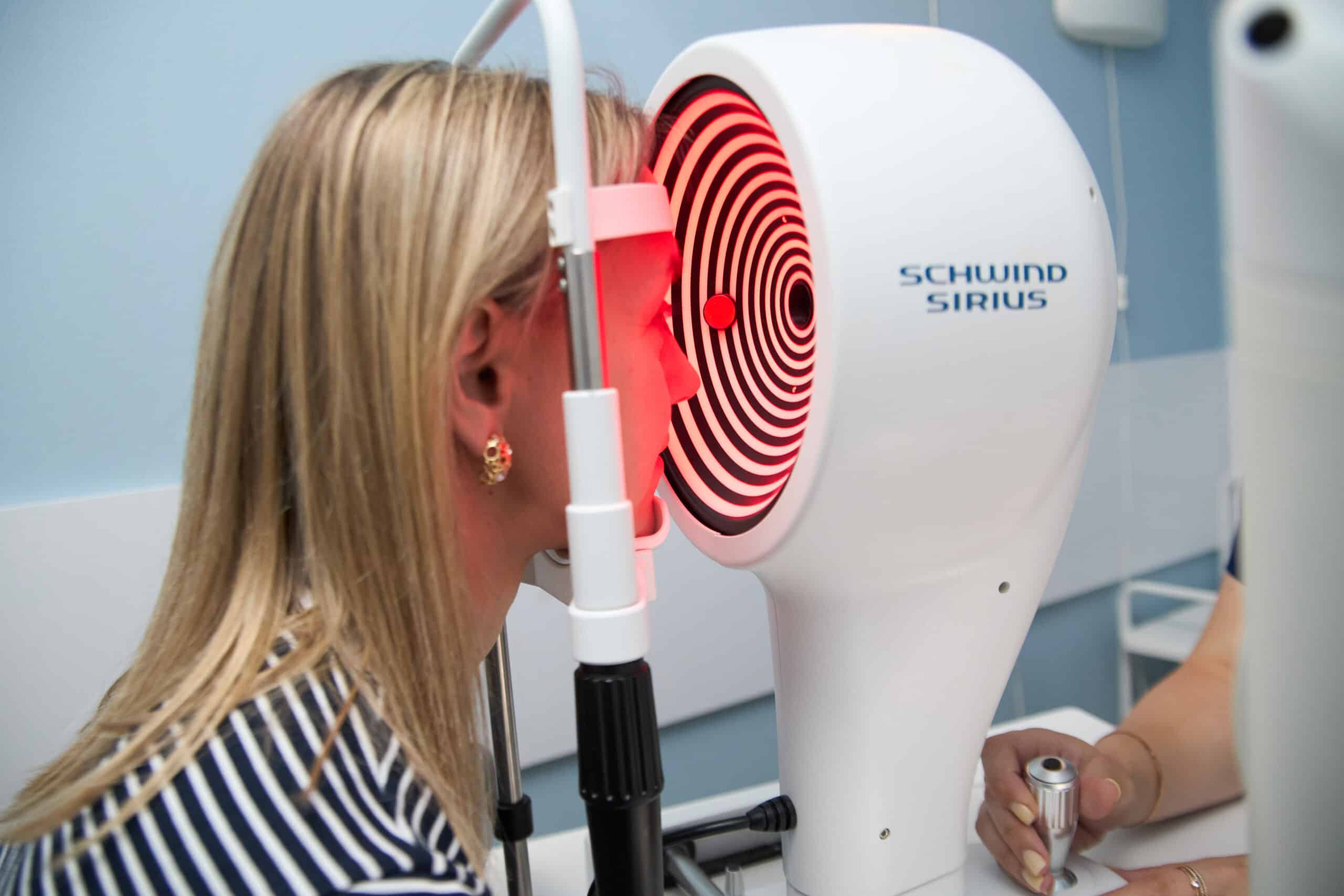
The device’s operation is based on the combination of two independent technologies within a single diagnostic platform: classic Placido-ring topography and a rotating Scheimpflug camera. The first technology maps the shape of the anterior corneal surface with high precision, while the second — by rotating around the eye’s optical axis — captures a series of cross-sections through the full thickness of the cornea and builds a three-dimensional model of it, including the posterior surface, which is inaccessible to conventional topographers.
During the examination at the Excimer Eye Clinic (Kyiv), the device performs a scan of the anterior eye segment in approximately one second, analyzing over 100,000 corneal points with a precision of around 1 μm. At the same time as topography, SCHWIND SIRIUS+ registers the wavefront reflected from the eye’s structures and compares it with an ideal flat wave — the same principle used by conventional aberrometers — providing a complete picture of the optical distortions of the entire visual system, not just the cornea.
By combining two diagnostic methods in one device, SCHWIND SIRIUS+ gives the ophthalmologist a substantially greater volume of data than a standard autorefractokeratometer or a separate topographer. In particular, the device determines:
- the precise corneal thickness (pachymetry) at any point;
- the elevation, curvature and refractive power of the anterior and posterior corneal surfaces;
- corneal diameter and deviations from normal shape, including early signs of keratoconus;
- displacement of the pupil center when transitioning from photopic to mesopic conditions (cyclotorsion), which is important for precise centration of the laser correction;
- static and dynamic pupillometry under different lighting conditions.
All the data obtained is combined into a clear color map of the anterior eye segment, which — much like the Acuity Map of conventional aberrometers — reflects the individual characteristics of a particular patient’s cornea, essentially its optical “fingerprint.” This map allows the physician not only to rule out contraindications to refractive surgery, but also to precisely calculate the extent and parameters of the procedure.
How SCHWIND SIRIUS+ Works
The device combines two cameras in a single housing. The first operates on the traditional principle — projecting Placido rings onto the cornea and registering their reflection to calculate a topographic map of the anterior surface. The second, a rotating Scheimpflug camera, rotates around the eye’s optical axis and captures a series of cross-sectional images of the anterior segment — from the anterior corneal surface to the posterior lens capsule. Dedicated software combines the data from both cameras, reconstructing a single three-dimensional model of the cornea and anterior eye segment, from which pachymetric, elevation and aberrometric maps are calculated.
What Can Affect Diagnostic Accuracy?
As with any optical measurement method, the accuracy of the results obtained with SCHWIND SIRIUS+ can be affected by tear film disorders: even with a normal cornea, an uneven tear film can distort topographic and wavefront readings. Significant opacities of the optical media, corneal scarring or advanced stages of keratoconus can make it difficult to build an accurate map. Pupil size also affects measurement accuracy — in cases of pronounced miosis, pharmacological pupil dilation may be required, since a narrow pupil limits the area available for analysis, and higher-order aberrations are most apparent with a dilated pupil.
Integration with the SCHWIND AMARIS EX500 Laser
The SCHWIND SIRIUS+ topographer operates within a unified SCHWIND diagnostic-and-surgical ecosystem together with the SCHWIND AMARIS EX500 excimer laser system, which is used for laser vision correction at the Excimer Eye Clinic (Kyiv). Since both devices are manufactured by the same company, topography, pachymetry and wavefront data obtained with SIRIUS+ can be transferred directly to the SCHWIND CAM (Custom Ablation Manager) treatment-planning software and used to calculate an individualized ablation profile — both for standard correction of myopia, hyperopia and astigmatism, and for personalized treatment based on the patient’s corneal or ocular wavefront (Corneal Wavefront / Ocular Wavefront Treatment).
The resulting individualized ablation profile is transferred to the AMARIS EX500 laser, which — with a pulse repetition rate of 500 Hz and an ultra-fine 0.54 mm laser spot — shapes the corneal surface to compensate for the measured optical distortions of the eye. Treatment precision is ensured by an active eye-tracking system, while the safety and stability of the outcome are supported by automatic pulse-energy regulation and protection of corneal tissue from excessive heating. This combination of highly precise SIRIUS+ topographic diagnostics and the modern AMARIS EX500 laser system ensures high accuracy, predictability and safety of laser vision correction.