


What is keratoconus?
The term keratoconus comes from two Greek words: “kerato” meaning “cornea” and “konos” meaning “cone”. Keratoconus is a degenerative eye disease in which the cornea becomes thin and conical due to structural changes, unlike the normal, spherical shape.
The essence of this condition is related to impaired optomechanical properties of the cornea’s basic substance. The fibers forming its base lose their strength characteristics. The cornea bulges forward exposed to intraocular pressure.
This pathology is typical for adolescents, but sometimes it may also be found in young people, more often up to the age of 30. Changes in the corneal shape occur slowly, usually over several years. However, there are also cases of more rapid keratoconus progression.
Why does keratoconus develop?
It is still unclear what causes keratoconus. Experts believe that the endocrine system, metabolic disorders, and connective tissue abnormalities have an effect. Moreover, when speaking about the causes of keratoconus development, it is worth mentioning the influence of heredity and unfavorable environmental factors (elevated radiation background and UV-irradiation may have a negative impact).
Risks of keratoconus
The conic-shaped cornea in keratoconus results in uneven refraction of light rays in different spots, which is why the visual acuity reduces (similarly to myopia). One sees distorted objects and crooked lines (similarly to astigmatism). In advanced keratoconus, corneal thinning (up to tearing) occurs, accompanied by a pronounced pain syndrome.
Signs of keratoconus:
- Inability to correct visual distortions with standard optical aids (glasses, contact lenses);
- Rapid myopia progression;
- Vision distortions similar to astigmatism;
- Halos and glare in the evening and at night;
- At the advanced keratoconus, a cone-shaped protrusion of the cornea is visible to the naked eye.
Stages of keratoconus
- The first stage is characterized by a slight corneal deformation.
- The second stage is characterized by increased corneal deformation and clouding of the upper part of the cornea.
- The third stage is characterized by a marked cone-shaped cornea, most of which becomes thin and opaque.
- In the fourth stage, the cornea visibly bulges, becomes almost completely thin and opaque.
As the disease advances, the visual acuity steadily decreases with increasing myopia and astigmatism. Painful corneal tears may occur, which cause vision to be lost almost completely.
Keratoconus diagnosis at Eximer Ophthalmology Clinic
Early keratoconus may be revealed only by a doctor when the necessary equipment is available. Early keratoconus is often mixed up with myopia or astigmatism, resulting in losing time and neglecting the disease. Therefore, it is worth remembering that sudden signs of myopia, its rapid progression, and distortions of vision similar to astigmatism can actually be symptoms of keratoconus development.
Eximer Ophthalmology Clinic employs a set of modern diagnostic equipment to examine the cornea in detail and detect keratoconus:
- Computerized corneal topography allows detecting the very first signs of keratoconus;
- Coherent tomograph can inspect the thickness of the cornea and analyze its condition without any contact;
- Endothelial microscope defines the quantitative and qualitative composition of the corneal endothelial layer of the eye.
Such diagnosis allows determining this condition in its earliest stages, guarantees the accuracy of the diagnosis and drawing a complete picture of the patient’s visual system, fixes and predicts the onset of the disease.
Ways to treat keratoconus
Depending on the condition’s stage, modern keratoconus treatment methods are used:
- Cross-linking;
- Stromal rings (segments) implantation);
- Keratoplasty.
Keratoconus treatment
Cross-linking
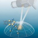
At the initial stage of keratoconus, a cross-linking procedure is performed. It involves reinforcing the cornea and stabilizing the keratoconus with a special laser. The procedure helps strengthen the corneal stroma, stabilize the progression of keratoconus, and stop the thinning of the cornea.
Stromal ring implantation
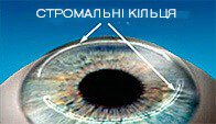
About one month after cross-linking, stromal rings are implanted into the cornea. The procedure gently changes the surface of the cornea, its refraction, and stabilizes the cornea. This procedure allows achieving good optical characteristics.
Keratoplasty

Keratoplasty is used with marked keratoconus. This corneal surgery is aimed at restoring the shape and function of the cornea and involves removing the damaged part of the cornea and replacing it with donor tissue.
Femtosecond laser to treat keratoconus
Today, it is possible to perform stromal ring implantation and keratoplasty using the most progressive and attenuated technique known as a femtosecond laser. The absence of mechanical impact shortens the recovery period, minimizes the risk of complications, allows for personalized intervention, and thus increases the effectiveness of keratoconus treatment.